Embryo in the Womb - by Leonardo Da Vinci
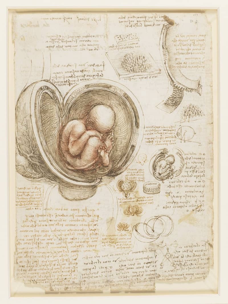
Leonardo da Vinci's embryological drawings of the fetus in the womb and his accompanying observational annotations are found in the third volume of his private notebooks. The drawings of Leonardo's embryological studies were conducted between the years 1510 - 1512 and were drawn with black and red chalk with some pen and ink wash on paper. These groundbreaking illustrations of the fetus reveal his advanced understanding of human development and demonstrate his role in the vanguard of embryology during the Renaissance. His famous embryological drawings of the fetus have since been collected and held in the Royal Collection at Windsor Castle in England.
Leonardo first ventured into human anatomy with the purpose of depicting the human body more accurately in his artwork. Although there is some evidence of his intentions to do so, Leonardo never published his work. The ultimate distribution of his journals and drawings is attributed to Orazio, the son of Francesco Melzi who was the faithful disciple to whom Leonardo entrusted his notebooks in his last testament. With the death of Francesco Melzi, Leonardo's life work was scattered and lost, never to be fully rediscovered.
In 1506 while in Milan, Leonardo's acquaintance with anatomist Marcantonio della Torre led him to many first-hand human dissections with the guidance of the younger professor. Four years later the expertise he gained with the help of della Torre would prove most useful in his studies of embryology. In one of his most famous drawings, Leonardo depicts a human fetus lying inside a dissected uterus. Leonardo is considered to be the very first in history to correctly depict the human fetus in its proper position within the womb. He was also the first to expertly draw the uterine artery and the vascular system of the cervix and vagina. Leonardo is credited with drawing the uterus with only one chamber, contradicting theories that the uterus was comprised of multiple chambers which many believed divided fetuses into separate compartments in the case of twins.
After his surgical exposure of a fetus within a cadaver, Leonardo's subsequent drawings portray an accurate understanding of the umbilical cord as consisting of vessels. Further drawings of the umbilical vessels illustrate his belief that menstrual blood nourished the fetus through the umbilical cord. Leonardo showed the umbilical cord connecting to the liver and his drawings of hepatic veins show the passing of blood to the heart. In his drawings, the feet of the fetus are crossed and the right foot is shown as blocking the urinary passage. Leonardo concluded that the position of the fetus's feet did not allow for the movement of urine through the urethra and so he theorized that the umbilical cord was the structure responsible for exporting the fetus's urine outside of the womb.
Leonardo used a method of cross-sectional representation for his depictions of veins, arteries, and nerves in order to show the layouts in greater detail. He was also fond of drawing four views of the subject so that every angle could be seen by the viewer for a more inclusive study, which he did for the drawings of the fetus. Leonardo's philosophy of the human body was often represented by comparisons to architecture. His drawings followed rigorous techniques often employed by architects to depict three-dimensional views of his subjects. He viewed the body as an architectural masterpiece created by nature, in which the skeleton was akin to rocks that laid the foundation for the body.
Leonardo expresses the human condition in a nutshell - indeed, his rendition of the womb resembles an opened horsechestnut casing. Inside is the beginning of us all laid bare. Five hundred years ago, this artist and scientist could portray the human mystery with a wonder that is not religious but biological he holds up humanity as a fact of nature.